Just amazing 💪🏾🤩 Team
Read More
What Do Chest X-Rays Show?
Posted: May 2nd, 2022 at 01:27PM
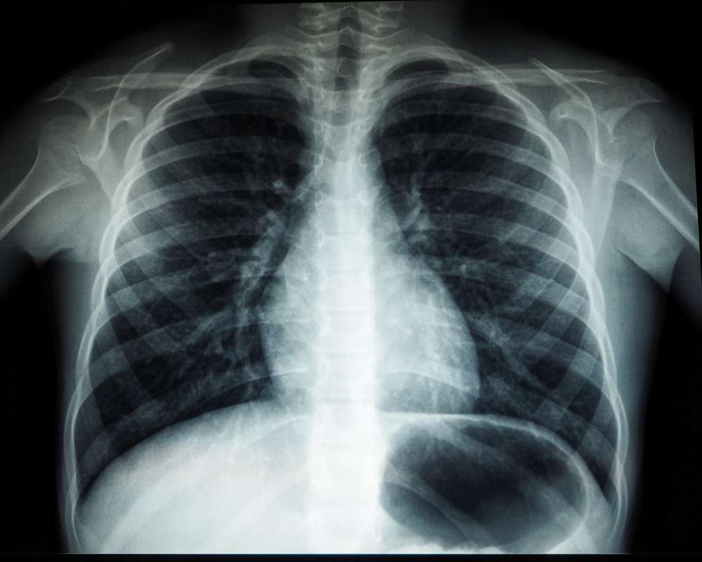
A chest x-ray is a common diagnostic imaging test that uses electromagnetic waves and small amounts of radiation. It provides doctors with information about the condition of the structures in your body. It can scan and create images of the organs and bones in the chest cavity.
A healthcare professional might prescribe a chest x-ray for various reasons. They're often used to screen for lung and heart problems, including tuberculosis, presence of fluid, tumors, and a variety of health conditions.
Chest x-rays have been a valuable imaging tool in the medical field for a long time because they can quickly provide physicians with diagnostic information about a patient's health and medical history.
If your doctor orders one for you, here are a few things you should know about what chest x-rays show and what you should expect during the procedure.
What Do Chest X-Rays Show?
A chest x-ray can create detailed images of the lungs, heart, and bones in the spine and chest. It helps detect and monitor treatment for a variety of respiratory illnesses and health conditions, such as:
- Pneumonia.
- Lung cancer.
- Emphysema.
- Congestive heart failure.
- Broken ribs.
- Infections.
Physicians also use chest x-rays to determine the condition and status of certain organs in your body. This includes:
- The health of your lungs and heart. Chest x-rays can provide your physician with detailed information about your lungs and heart's size, shape, and condition. They help in the fastest and easiest detection of abnormalities or fluid buildup around the heart, collapsed lungs, pneumonia, cancer, emphysema, cystic fibrosis, and other respiratory ailments.
- Calcium deposits. Calcium in the blood vessels or heart can indicate weak heart muscles or valves, as well as increased chances of a heart attack.
- The placement of a pacemaker or other medical devices. Chest x-rays can check the performance and position of pacemakers and defibrillators (equipment that maintains a person's heart rhythm), and catheters (tubes that are utilized to deliver drugs or receive dialysis).

When Should You Get a Chest X-Ray?
Your physician may order a chest x-ray if you have certain symptoms that could be caused by problems with your lungs or heart.
These symptoms might include
- Coughing up blood.
- Chest pain.
- Difficulty breathing.
- Fever.
- Persistent coughing.
How Do Chest X-Rays Work?
The x-ray machine creates a small amount of ionizing radiation that goes through your body to produce an image. The x-ray scan is recorded on film or a digital memory device.
The denser structures in your body (such as bone) absorb more of the x-ray beam than the less dense tissues (such as lungs). As a result, bones appear white, and soft tissues appear gray in the final image.
How Do You Prepare for a Chest X-Ray?
Most often, a chest x-ray is done as an outpatient procedure. That means you can go home the same day. You also won't need any special preparations for a chest x-ray.
What Should You Expect During a Chest X-Ray?
A chest x-ray is an easy, non-invasive, and painless test. You will be asked to remove any clothing, jewelry, piercings, or other objects that might get in the way of the x-ray. You will be given a lead apron to reduce exposure and protect your body against radiation (although the amount of radiation you'll be exposed to during the procedure is very low).
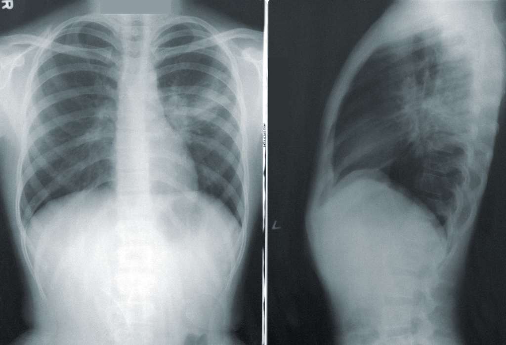
You'll be asked to stand or sit in front of the x-ray machine by a radiology technologist. A chest x-ray may involve two parts:
First, to produce a standard chest x-ray image (posteroanterior view) showing the front of your chest, the technologist will position you and the x-ray machine so that your chest is centered on the radiographic plate, with your hands on your hips.
Next, you may be instructed to stand sideways to the metal plate of the X-ray machine with your arms elevated. This position produces a lateral chest view or a side view of your chest and is performed to obtain further information.
You may be asked to stand very still or hold your breath at times during the procedure, which only takes a few minutes.
 Once the chest x-ray is complete, the technologist will review the images to make sure they're clear. If not, more pictures may need to be taken. The technologist will then process the film and send it to a radiologist (a doctor who specializes in comprehending x-rays) for interpretation.
The radiologist will look for anything that appears abnormal on the film and send a report to your physician, who will discuss the results with you after comparing images from your x-rays with previous scans if you've had any. The results of your chest x-ray will help guide treatment decisions.
What are the Risks of Getting a Chest X-Ray?
There is minimal risk involved in having a chest x-ray. X-ray machines use a small amount of ionizing radiation, which has the potential to cause cancer. However, many physicians agree that the benefits of getting an x-ray vastly outweigh this small risk. Additionally, x-rays are constantly regulated and monitored to ensure that the radiation levels required to produce images are always set to a minimum.
If you're pregnant or think you may be pregnant, it's important to let your doctor know before agreeing to have a chest x-ray, so the technologist can take proper precautions.
When Do You Receive the Results of Your Chest X-Ray?
Your doctor will go over the results of your x-ray as soon as they're available, which is usually within one to two days. If there are any urgent findings, the radiologist will likely contact your doctor immediately. If everything looks normal, you'll probably get a call from your doctor's office saying that the results were negative and that no follow-up is needed.
What Happens After You Receive Abnormal Results?
While a chest x-ray can reveal many health conditions, an abnormality found in your results doesn't necessarily mean that you have a respiratory ailment. There are many possible causes of an abnormal result, and your doctor will likely order additional tests to determine the cause. This could include a CT scan, which provides more detailed images of the lungs, or a bronchoscopy, which involves inserting a small camera into the lungs to get a closer look.
If you think you might need a chest x-ray, contact e7 Health today or book an appointment online.

Plenty of services to be used. Easy to use and quickness. I was in and out.
Read More
Professional and warm very rare these days.
Read More
I had a great experience here. I was waiting less than 3 minutes to be seen, and I received all of my results within a few hours!!
Read More
Excellent 10 to 15 Minutes Done my Appointment
Read More
Fun!
Read More
Very Nice people. They kept their word on privacy and stood on business. I never had to take a urine test while being on medication to help me get off opiates. I showed proof what medication I was on and the proces went smooth.
Read More
My nurse/aide made the entire experience delightful! Was there for a urinalysis but was bladder shy for some reason even after two bottles of water.. she was upbeat, kind and so helpful. She made the experience wonderful instead of stressful that's it easily could have been. The facility is extremely clean and the staff is top notch from beginning to end! It's completely across town from me but I will use this facility from now on.
Read More